


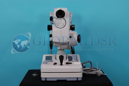
US $33,995.00
| Condition | Seller refurbished
:
An item that has been restored to working order by the eBay seller or a third party not approved by the manufacturer. This means the item has been inspected, cleaned, and repaired to full working order and is in excellent condition. This item may or may not be in original packaging. See the seller’s listing for full details.
|
| Seller Notes | “Near flawless condition, hardly ever used.” |
Directions
Similar products from Retinal Digital Cameras
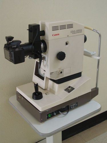
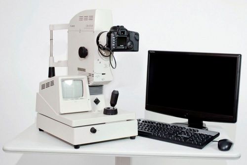
Pre-owned Canon CR4-45NM Retinal Camera upgraded to digital
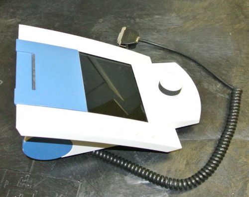
OCULUS visual field analyzer perimeter control box
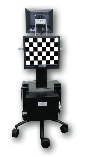
Zeiss Humphrey Instruments 740 Field Analyzer
Kowa Nonmyd a-D Retinal Fundus Camera Non-Mydriatic
Zeiss Humphrey 750 HFA-II Visual Field Refurbished Warranty Printer & Table, GPA
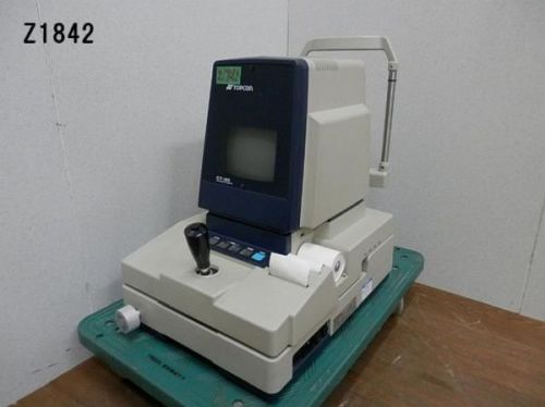
Topcon TRC-NW6S Retinal Camera
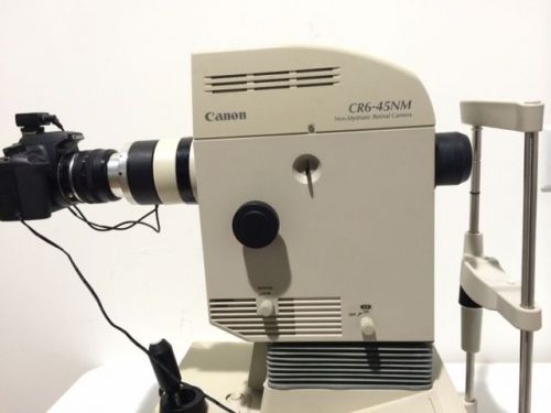
Canon CR 6 - 45 NM Digital Non-Mydriatic Retinal Camera/Fundus Camera
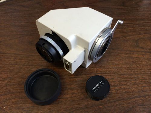
Topcon 1X Relay Lens, for TRC-50X OR 50IA Fundus. USED TO CONVERT TO DIGITAL.
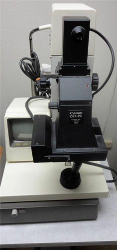
Retinal Camera - Non-Mydriatic Canon CR4-45mm model # 205206
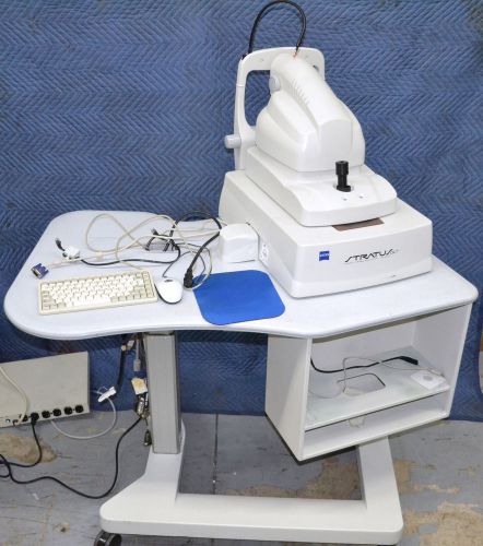
Zeiss OCT 3000 Tomographer Glaucoma Retinal with Power Table
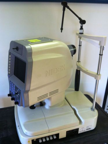
NIDEK NM-1000 Non-Mydriatic Fundus Camera Mfg 2006 Excellent Condition Retinal
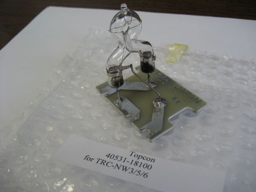
Topcon Xenon Flash Tube # 40531-18100 for Topcon TRC-NW3/5S/5SF/6/6S, GREAT FIND
Canon CR-6 Non-Mydriatic Retinal Fundus Camera w Synamed Digital System
Nidek NM 1000 DIGITAL IMAGING NON-MYDRIATIC FUNDUS CAMERA
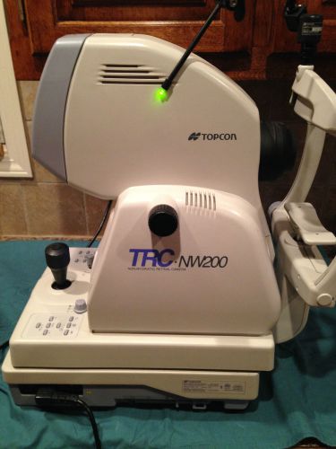
TOPCON TRC NW200 NON MYDRIATIC RETINAL CAMERA
Zeiss Humphrey 740i Visual Field Analyzer
Zeiss Humphrey 750 Visual Field Perimeter Analyzer With Software & Powered Table
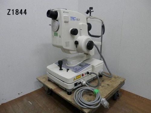
Topcon TRC-50DX Mydriatic Retinal Fundus Camera
People who viewed this item also vieved
INFUMED Lindstrom Spatula FOR USE IN OPHTHALMIC SURGERY TITANIUM
new low priceSchepens Scleral Depressor medium for ophthalmic surgery instrument
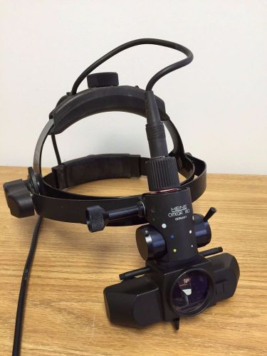
Heine Indirect Ophthalmoscope, model Omega 180 with Carl Zeiss 6v power supply.
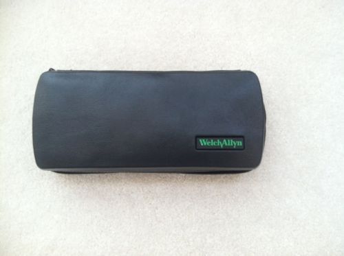
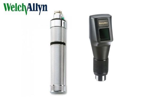
Welch Allyn Panoptic Ophthalmoscope Hard Carrying Case Model 05258 M
WELCH ALLYN 3.5V STREAK RETINOSCOPE WITH DRY BATTERY HANDLE-FREE SHIPPING

WELCH ALLYN 3.5V STREAK RETINOSCOPE WITH "C" BATTERY HANDLE-FREE SHIPPING

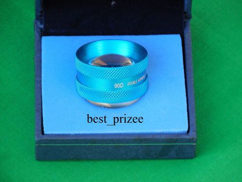
ophthalmology lens 90D aspheric lens
By clicking "Accept All Cookies", you agree to the storing of cookies on your device to enhance site navigation, analyze site usage, and assist in our marketing efforts.
Accept All Cookies