US $2500
Directions
Similar products from Measuring Devices
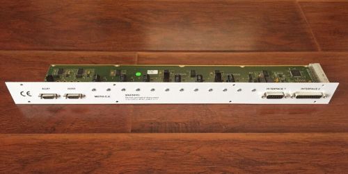
CTC Analytics Motio E.K Board Autosampler Dionex PN VK-AB8954
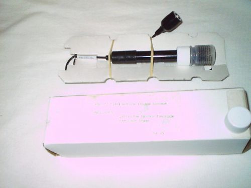
YSI 112-1 PH ELECTRODE,DOUBLE JUNCTION
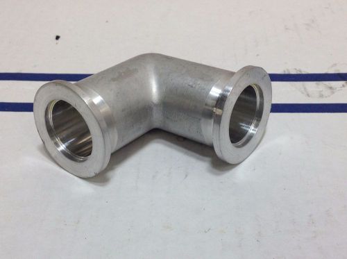
High Vacuum Elbow DN 25 ISO KF Aluminum
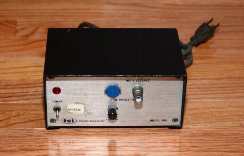
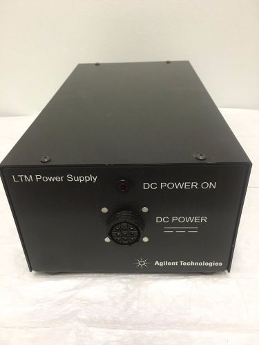
Thermo Systems Inc TSI Photomultiplier Power Supply - Model 965
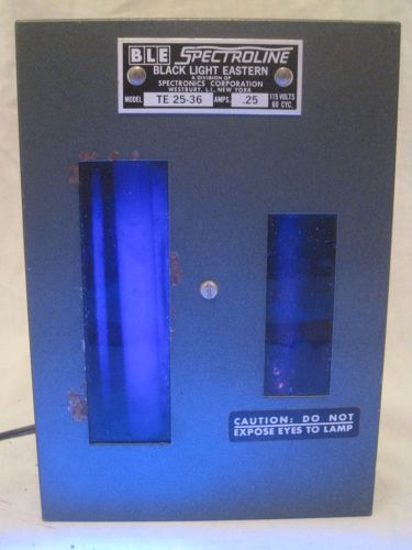
vintage BLE Spectroline Black Light Eastern TE 25-36 dual lamp Spectronics
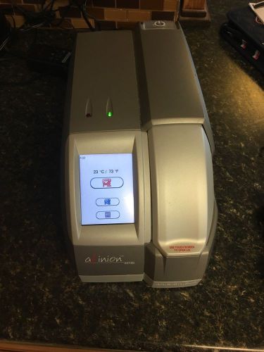
Axis-Shield Afinion AS100 Analyzer PoC Diabetes w/ AC adapter
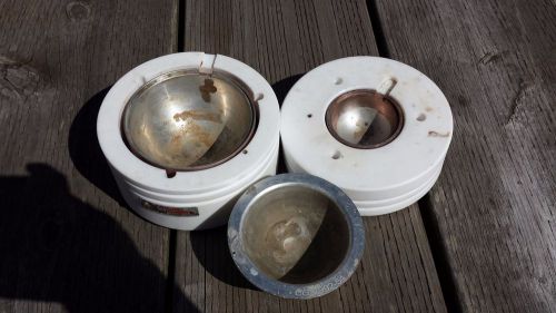
OPTITHERM reaction blocks for 250ml and 100 ml RB flasks
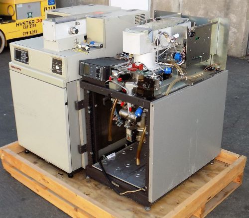
TWO HP HEWLETT PACKARD 5989B MASS SPECTROMETERS
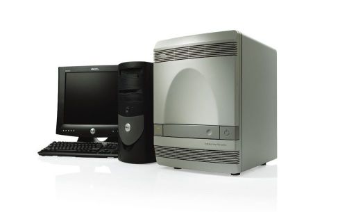
Applied Biosystems ABI 7300 Real Time PCR w/ PC & Software
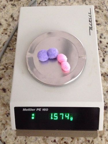
METTLER PE 160 LABORATORY SCALE
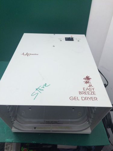
HOEFER EASY BREEZE GEL DRYER Model SE 1200 With Extra Rack
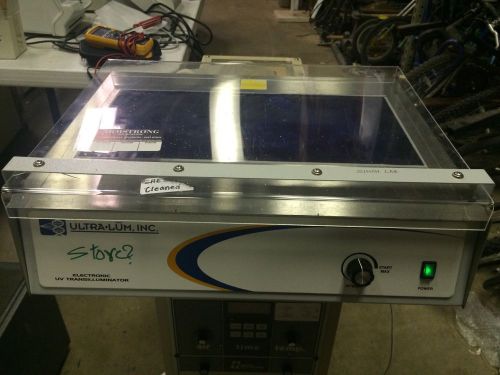
Ultra.Lum Inc 900-1425-05 EABC-20 Electronic Multiwave Transilluminator
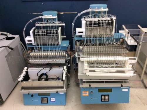
Lot of 2 Brandel Suprafusion Tissue Perfusion Systems SF-2518 & SF-2520
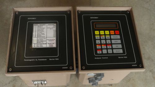
Servomex Sybron Analyser Control 1101 + Paramagnetic O2 Transducer 1131
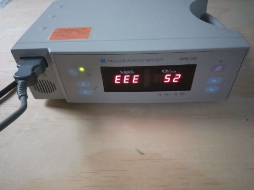
Nellcor Puritan Bennett NPB-290 with Nellcor SpO2 Cable
Westinghuse hollow cathode Lamp Be type WL 36013 BE 21R2
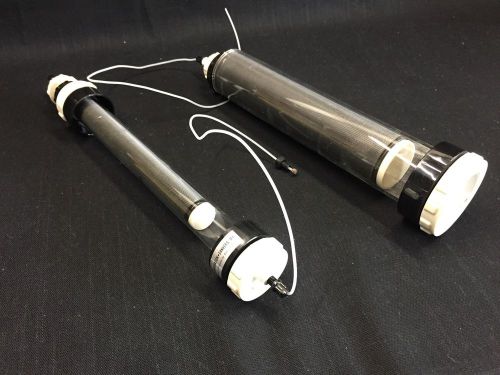
(Lot of 2)Amicon FPLC Chromatography Columns
Agilent G1947 A APCI Atmospheric Pressure Chemical Ionization Source
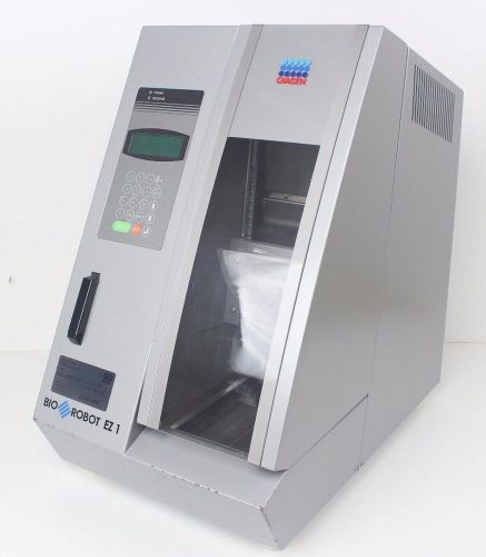
Qiagen EZ1 Workstation Liquid Handler w/ DNA Bacteria Protocol Card
People who viewed this item also vieved
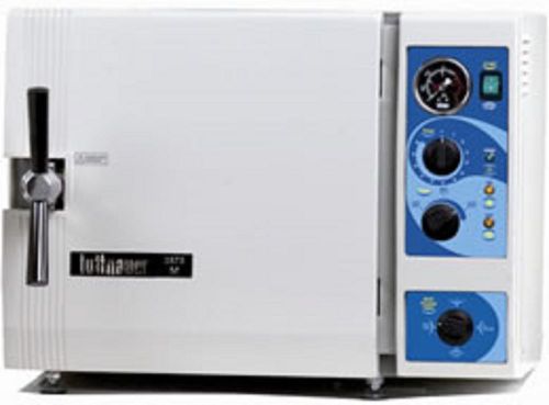
Brand NEW Tuttnauer 3870M - Large Capacity Manual Autoclave
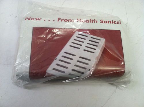
Health sonics ASEP Instrument caddy
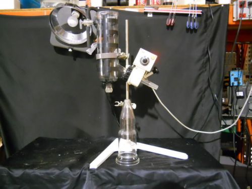
Buchi Rotavapor Evaporator Missing some glassware Type KRv 65/45
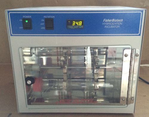
FisherBiotech Hybridization Incubator Oven Model FBHI10 Fisher Biotech FBH110
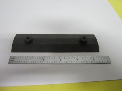
GRAPHITE PLATE FROM ION IMPLANTER VERY NICE LASER OPTICS AS IS BIN#54-02
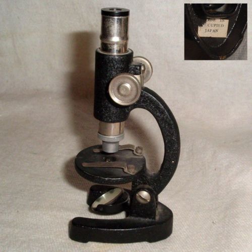
60yr OCCUPIED JAPAN black & chrome MICROSCOPE w/MIRROR no damage works +1 free
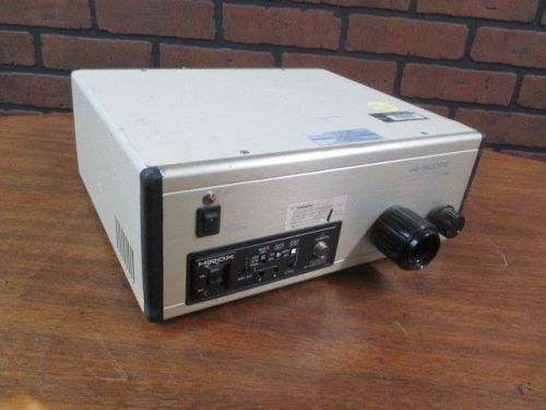
HiRox Hi-Scope KH-2200 MD3 Compact Micro Vision for Video Microscope - Warranty
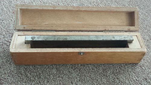
Hacker Instruments Inc H/I Microtome Knife Blade 180mm 7 in. Sideways profile C
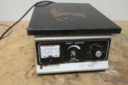
LABLINE ORBITAL SHAKER MODEL 3520
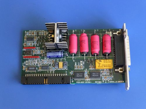
Newport 25792 Plug-in Driver Card for ESP300 Motion Controller
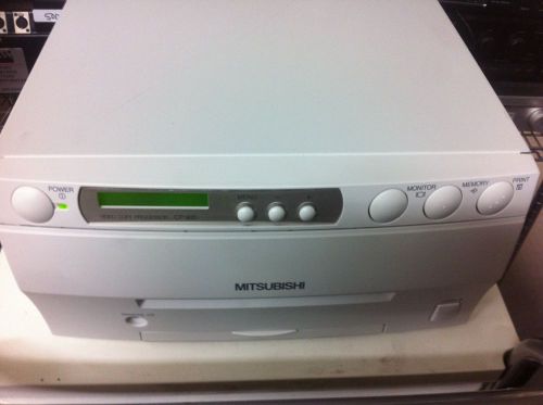
MITSUBISHI COLOR VIDEO COPY PROCESSOR ( MODEL CP-900UM )
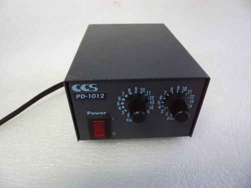
PD-1012 CCS Box Vision Light Power Supply
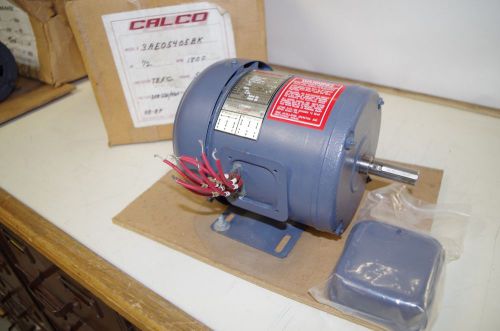
CALCO 1/2HP AC MOTOR # 3AE05405BK 208-230/460VAC 60HZ. 1720RPM
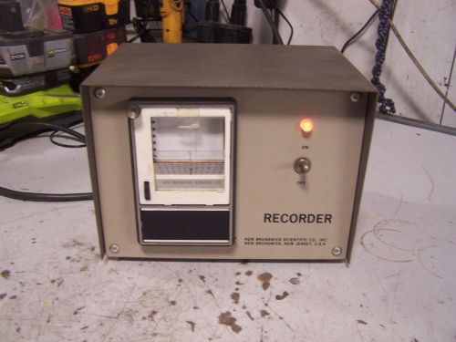
NEW BRUNSWICK M1055-7701 SCIENTIFIC RECORDER 115 VAC 1 PHASE .5 AMPS
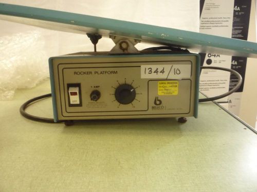
BELLCO TECHNOLOGY ROCKER PLATFORM (ITEM # 1344/10 )
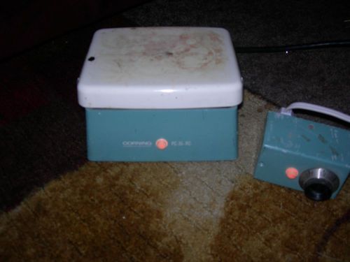
CORNING - PC-35-RC REMOTE CONTROLLED LAB HOT PLATE/STIRRER
By clicking "Accept All Cookies", you agree to the storing of cookies on your device to enhance site navigation, analyze site usage, and assist in our marketing efforts.
Accept All Cookies