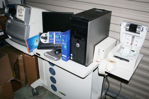
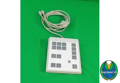
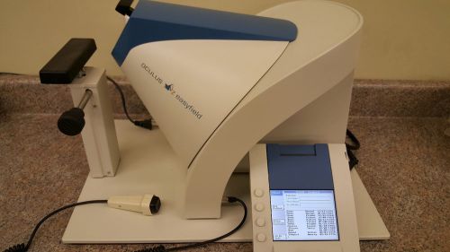
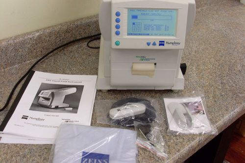
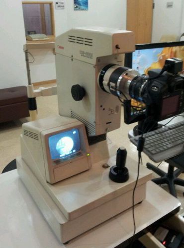
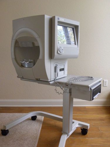
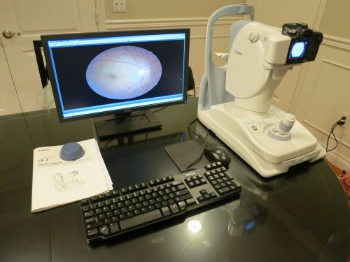
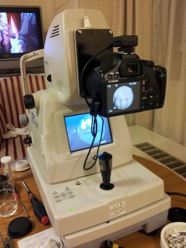
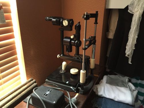
Marco Talia RTA-5 OCT Fundus Camera Macula RNFL ONH DISC & Fundus Imaging Marco Talia RTA-5 Unit is working to factory specifications. The device comes complete, patient ready, with the following items: Talia RTA-5 SLO Marco Talia table Dell Optiplex 745 PC Printer Monitor, Keyboard, & Mouse Instruction booklet Manufacturers Description: The RTA 5 represents a new perspective in diagnostic imaging. It is the first system which integrates high speed scanning laser ophthalmoscopy (SLO) with high resolution digital fundus imaging for early detection, diagnosis, and progression analysis of pathology involving the macula, perimacular regions, optic nerve head, and peripapillary regions of the retina. RTA 5 Benefit Summary - The highly versatile RTA 5 delivers comprehensive diagnosis of glaucoma, diabetic retinopathy, age related macular degeneration, and many other retinal pathologies – with speed, accuracy, and an intuitive user interface. Patients and practices benefit from RTA 5 capabilities, such as: • Up to 72° x 60° digital fundus Images at 9.4 mega pixel resolution • Actual SLO, B-scan slit images less than 0.7 degrees apart • Retinal thickness analysis at 1,400 - 3,200 data points • 2D and 3D retinal thickness maps • Dynamic 3D Anatomy Imaging through all scan volumes at real slit sections • Retinal nerve fiber layer (RNFL) cross-section charts with temporal-to-temporal TSNIT Analysis • Peripapillary and optic nerve head (ONH) analysis with rim/cup measurement • Automated progression analysis with deviation probability reporting • Non-mydriatic capability (with 3+mm pupils) • Remote viewer software for analysis and patient consultations • EMR compatible Comprehensive Fundus Image Acquisition - In 0.33 - 0.48 seconds, the RTA 5 simultaneously captures high resolution digital fundus images with each series of SLO scans of the full posterior pole. The result is a 72° x 60° Wide Field or 60° x 40° Standard Field image, at up to 9.4 mega pixel resolution. Automatic Registration of Fundus Images - Utilizing a proprietary registration algorithm, the RTA 5 automatically aligns each image based on the patient’s vascular patterns for enhanced accuracy and repeatability. This feature ensures reliable follow-up examination results without full dependence on consistent patient fixation. High Speed Scanning - The RTA 5 Standard examination includes three scans of the macula, each composed of twenty-four slit images per 20° x 10° area, plus one 10° x 10° scan of the ONH/disc composed of sixteen slit images. Scan times are just 0.48 seconds (macula) or 0.33 (ONH) and are essentially unaffected by eye movements. The collective 88 min./200 max. slits are separated by an average of 0.7 degrees to generate 1,408 min./3,200 max. real retinal thickness samples. Such scan density ensures minimum interpolation of data. Broad scanning areas minimize examination time, reduce the number of composite retinal images and enhance overall data integrity. Within moments, exceptional photo documentation and quantitative results are available for viewing, printing, and sharing with patients and other clinicians. SLO Slit Images, Retinal Thickness, and Topography - The RTA 5 provides an automatic presentation of all actual, individual slit images within a given scan. Slit images cross-section the retina along a tight vertical grid making it possible to evaluate false-positive artifact and identify real pathology during an examination. Additionally, a quality assessment (QA) score reflects SLO slit integrity for increased user confidence in the validity of the results. Quantitative Stereometric Measurement - The RTA 5 projects narrow(solid state 532 nm), laser light onto and through the retina, harvesting reflectance between the vitreoretinal and chorioretinal interfaces. A thickness algorithm identifies the location of the anterior (RNFL) and posterior (RPE) retinal borders where the laser light is fully absorbed. Average retinal thickness is determined at sixteen points along each vertical slit. The RTA 5 combines 25,000 A-scans in the final B-scan slit images – presented in color and grayscale modes. This unique density of data provides more accurate analysis of thickness changes and pathology structure across the macula and ONH. Disclaimer: The sale of this item may be subject to regulation by the U.S. Food and Drug Administration and state and local regulatory agencies. If so, do not purchase this item unless you are an authorized purchaser. If the item is subject to FDA regulation, I will verify your status as an authorized purchaser of this item before shipping of the item. If you have questions about legal obligations regarding sales of medical devices, you should consult with the FDA's Center for Devices and Radiological Health: http://www.marco.com/brochures/RTA5%20Brochure.pdf
By clicking "Accept All Cookies", you agree to the storing of cookies on your device to enhance site navigation, analyze site usage, and assist in our marketing efforts.