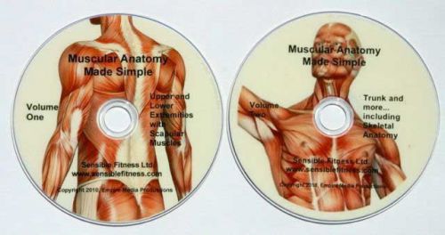
Our two-disc DVD series gives you a detailed muscle anatomy lesson including the origin, insertion, and action of spotlighted muscles using both layman's terms and the proper medical terminology. In addition to learning the muscles of the human body, you will learn the impact a double-jointed muscle has on the human body, the etiology (cause) of various musculoskeletal injuries, along with pathology related to the musculoskeletal system. We make a very complicated topic easy to understand in this human anatomy and physiology resource. Free sample video footage is available by searching YouTube for "Sensible Fitness Anatomy". Muscle anatomy of the human body included: Volume One. Lower and Upper Extremities Including Scapular Muscle Anatomy Triceps. Detailed anatomy of the three muscles that make up the back of your arm. Learn which head is a double jointed muscle and how that affects motion. Biceps. Even children know how to pose this muscle, now learn the anatomy and actions. Deltoids. The muscular anatomy of the anterior, lateral, and posterior head of this muscle is detailed. Pectoralis Major. This muscle makes up the bulk of your chest and is responsible for many functional and sports related actions. Trapezius. This diamond shaped muscle's anatomy is broken down into the upper, middle, and lower trapezius, with the actions of each discussed. Latissimus Dorsi. The largest of your back muscles, and one that is responsible for many actions with the shoulder, spine, and even the pelvis. Rhomboids. Important for good posture, the rhomboid anatomy is explained in detail. Levator Scapulae. This muscle and often be a pain in the neck. Literally. Going from the scapula up to the base of the skull, knowledge of the anatomy of the Levator Scapulae is crucial for various types of therapist's. Pectinius. The most proximal, and smallest of the inner thigh muscles. Adductor Brevis. Very few know that there are 5 muscles that make up the "inner thigh" muscles. Learn them all with this DVD. Adductor Longus. After watching this DVD, your knowledge of the hip adductor anatomy will surpass that of your peers. Adductor Magnus. As with many muscles of this complex, Adductor Magnus gives us other motions than just hip adduction. Learn what they are! Gracilis. This, the longest of the hip adductors is a double jointed muscle, as it crosses the knee. Learn the relevance of this and how it relates to movement. Gluteus Maximus. The largest and most powerful of your "butt" muscles. The anatomy is detailed, along with the multiple actions this muscles gives the human body. Vastus Intermedius. The deepest of the quadriceps, not seen from the surface. Vastus Lateralis. This muscle can become stronger than the Vastus Medialis, which causes knee dysfunction. Learn more about this! Vastus Medialis. Learn the anatomy of this muscle and the importance of strengthening it for a healthy knee. Rectus Femoris. The only double jointed muscle of the quadriceps group, that also has action on the hip. Semitendonosis. This is one of the hamstring muscles and shares a common union with another hamstring muscle. Semimembranosis. The hamstrings give humans movement at the knee, and the hip. Learn the muscular anatomy and see the motions these muscles provide. Biceps Femoris. The third, and most lateral muscle of the hamstring complex. Gastrocnemius. This calf muscle also crosses the knee, giving us knee flexion. See the detailed anatomy. Soleus. A deep, but very important muscle of the calves. Volume 2: Human Core Muscles and more... including Skeletal AnatomyRectus Abdominis. The almighty abs! They are more than just a "six pack". Learn the muscle anatomy for this powerful stabilizer of the spine. Internal Obliques. Sometimes forgotten because it lies deep, this muscle has a number of important actions. External Obliques. The anatomy of this muscle differs from the deeper internal oblique. Learn how! Transversus Abdominis. This could be one of the most important core muscles for a healthy back. Spinal Erectors. Some muscles in this group are very small, while some travel nearly the length of your entire spine. All are important and give our body unique movement. Quadratus Lumborum. Seeing the anatomy of this muscle will give you a clearer picture of how it impacts the movement of our spine. Gluteus Minimus. This is a very deep muscle that contributes to our "butts". Gluteus Medius. This muscle has a huge impact on our gait as humans, and is often impacted greatly with hip surgeries. Learn how! Illiacus. Learn where this muscle attaches to the skeleton, and the movement it gives us. Psoas Major. This muscle connects to the spine, which can sometimes be a concern. Learn why. Piriformis. The muscular anatomy will reveal how this muscle can cause "sciatica". Sartorious. This is the longest muscle in the human body. Serratus Anterior. A very important muscle for the health of your shoulder. Many do not understand the anatomy, but we make it easy. Tensor Fascia Latae. Learn the origin, insertion, and action of this human muscle. Supraspinatus. The most common muscle of the rotator cuff to cause problems. We get into much detail with the anatomy of this one. Infraspinatus. An important external rotator of the shoulder joint. Teres Minor. Learn how a strong rotator cuff will allow overhead motions without problems. Subscapularis. The only internal rotator of the rotator cuff group. Flexor Carpi Radialis. Learn the anatomy of this wrist flexor. Flexor Carpi Ulnaris. Learn where this muscle attaches and it's link to "golfer's elbow". Extensor Carpi Ulnaris. Lateral epicondylitis is discussed with the wrist extensors anatomy. Extensor Carpi Radialis Brevis. This is the shorter of the wrist extensors that are discussed. Extensor Carpi Radialis Longus. Learning the anatomy of this muscle will help therapists and trainers a great deal. Human skeletal anatomy included: Clavicle. Commonly called your "collar bone". Learn what muscles attach to it. Femur. The largest bone in the human body. Fibula. A long thin bone that goes down the outside of your shin. Humerus. A nerve gets pinched against this bone when you hit your "funny bone". Patella. A floating bone, called the knee-cap by many. Illium. The largest of the three bones that make your pelvis. Ischium. Your hamstring muscle group originates here. Pubis. This part of your pelvis helps identify a female vs. a male skeleton. Learn how. Radius. One of two bones that make your forearm. Ribs. There are a number of different types of ribs. Learn more with this anatomy lesson. Sacrum. This is often called your tail bone. Coccyx. A small bone at the bottom of your sacrum. A number of muscles attach to this bone. Scapula. A lot of muscle attach to this nearly free-floating bone, to keep it stable. Sternum. This bone is made up of the Manubrium, Gladiolus, and Xiphoid Process. Tibia. Ever crack your shin and wince in pain? This is the bone that was hit. Ulna. This bone makes up most of your wrist. Vertebrae (included with Spinal Erectors anatomy). We visit about the vertebrae and also discuss the intervertebral discs. Herniated discs included in this anatomy lesson.
By clicking "Accept All Cookies", you agree to the storing of cookies on your device to enhance site navigation, analyze site usage, and assist in our marketing efforts.