

US $7,599.00
| Condition: |
Used: An item that has been used previously. The item may have some signs of cosmetic wear, but is fully
operational and functions as intended. This item may be a floor model or store return that has been used. See the seller’s listing for full details and description of any imperfections.
...
|
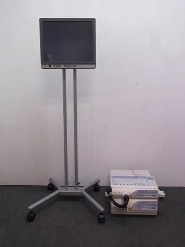
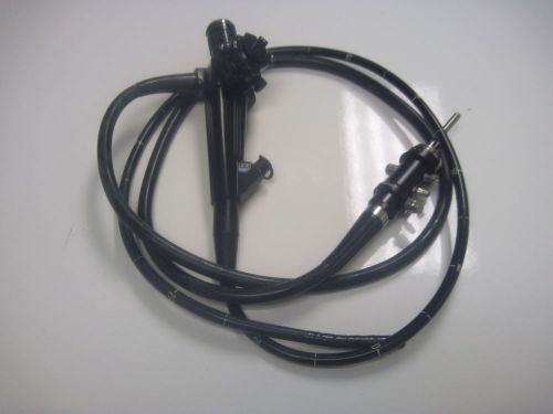
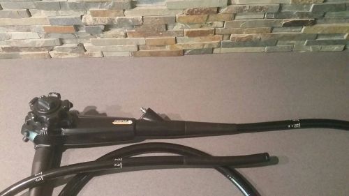
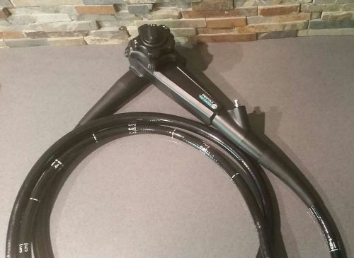
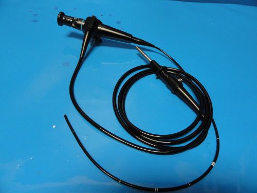
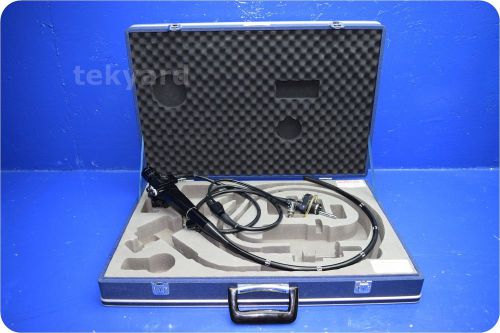
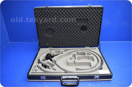
Brand | Olympus |
| Country/Region of Manufacture | Japan | ||
| Model | CLV 260 CV 260 ELVIS LUCERA |
Directions
Similar products from Other Equipment for Endoscopy & Laparoscopy
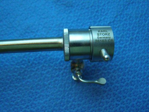
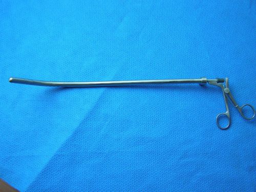
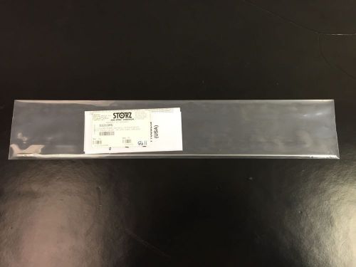
1:Storz 26163 CR Hysteroscope Operating Sheath Endoscopy laparoscopy Instrument
1:Unit KARL STORZ 26600BB Monopolar Hook Laproscoopy Instruments.
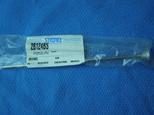
1/ Karl Storz 28124BS OBTURATOR SHARP Reusable Endoscopy Instruments
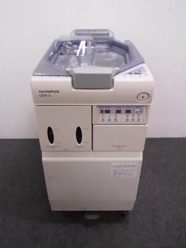
OLYMPUS ENDO REPROCESSING SYSTEM OER-2
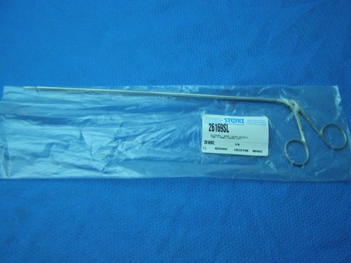
1:STORZ 26169SL Micro Dissect Scissors CVD Laparoscopy Endoscopy Instruments
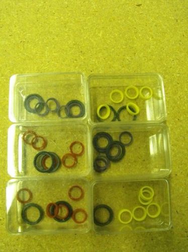
OLYMPUS O-RINGS SEALS 6 SMALL CONTAINERS 5 PACKS MAJ-690 CAPS 5829500 NEW
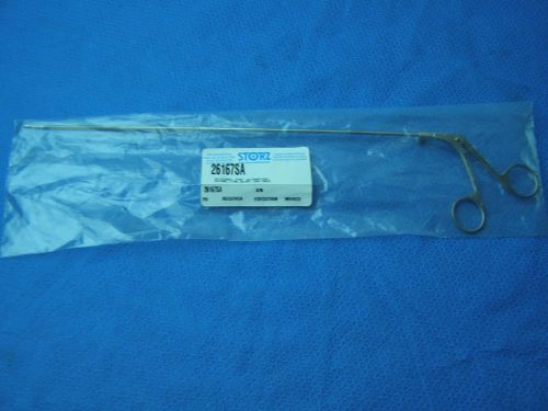
1:STORZ 26167SA Scissors UltraMicro Suture Fine Blunt 3mm Endoscopy Instruments
1/ Karl Storz 27021 O OBTURATOR Probe 5mm Blunt Reusable Endoscopy Instruments
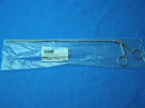
1:STORZ 26169 SMF Scissors Laparoforce HOOK Endoscopy Instruments
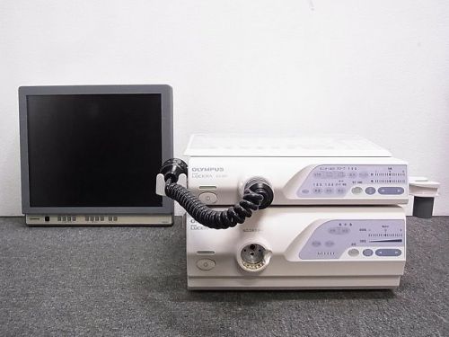
OLYMPUS CLV 260 CV 260 ENDO ELVIS LUCERA
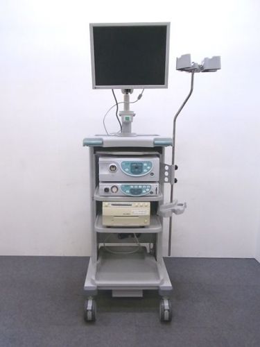
Video Endoscope System 4400 HD FUJI FILM
Olympus Laparoscopic Rigid 10mm CURVED Shaft Cup Forceps A5584 Lot Of 1
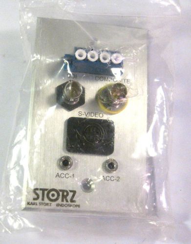
Storz Endoskope RGBO panel w/ SDI, composite, s-video, ACC-1 + ACC-2 ports
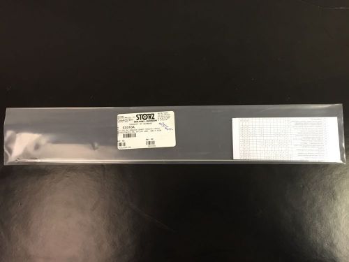
Karl Storz 33310A Clickline Babcock Grasping Forceps Insert Fens 5mm x 36cm
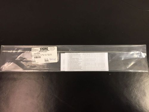
Karl Storz 33310CG Clickline Cone Serrated Tip Grasping Forceps Insert Doubl Cup
Karl Storz 33310MS Clickline Manhes Duckbill Dissecting Grasping Forceps Insert

Karl Storz 33300M Clickline Metal Outer Tube 5mm x 36cm w/ Luer-Lock
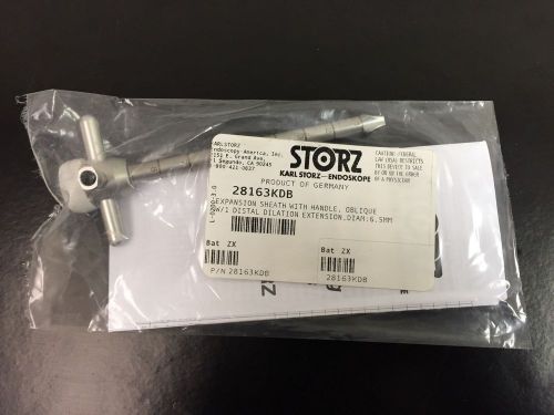
Karl Storz 28163KDB Expansion Sheath w/ Handle Oblique w/ 1 Distal Dilation Extn
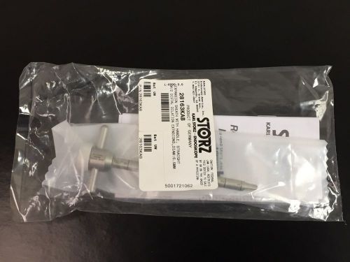
Karl Storz 28163KAB Expansion Sheath w/ Handle Straight w/ 2 Distal Dilation Ext
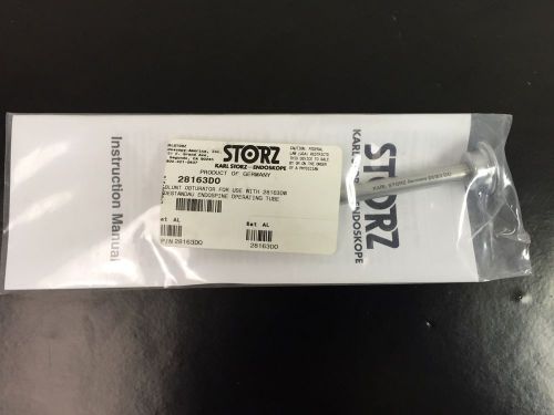
Karl Storz 28163DO Blunt Obturator f/ 28163DW Destandau Endospine Operating Tube
People who viewed this item also vieved
Olympus JF-10 Fiber Optic scope
Pentax EC-3872LK Colonoscope Endoscope OEM Flexible Endoscopy
Pentax EG-3470K Gastroscope Endoscope endoscopy OEM
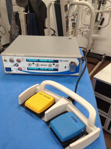
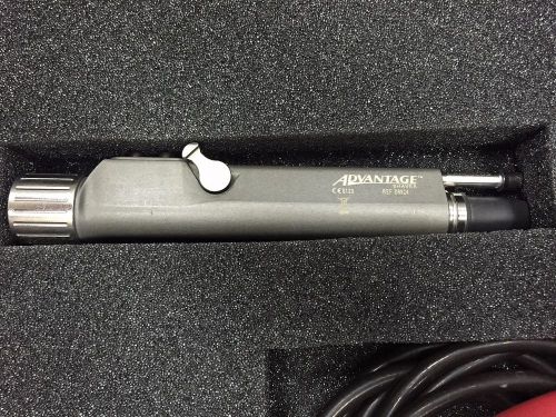
Conmed Linvatec Hall Advantage Driver D3000 Shaver Power System w/footswitch
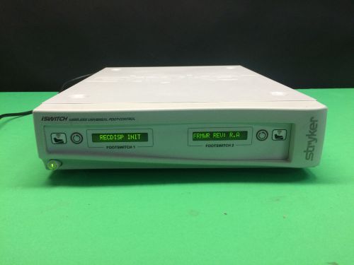
Stryker iSwitch Wireless Universal Foot-Controller 277-200-100
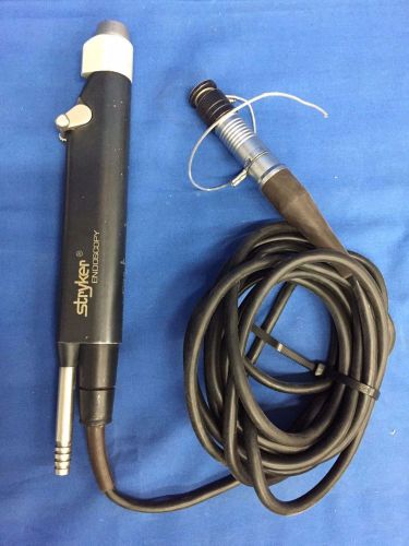
ConMed Linvatec D9824 2-Button Shaver
Stryker Endoscopy Model No. 275-701-500 12K Shaver
Karl Storz 30107LP Trocar and Cannula

Karl Storz 30107LP Trocar and Cannula. NEW.
Zimmer 60-3953-034 Arthoscope Trocar
Pentax OF-B121 Air/Water Valve Videoscope 30K, 40K, 90K, 90i & Fiberscope V & W
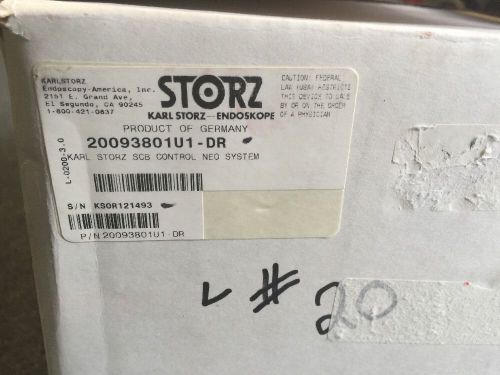
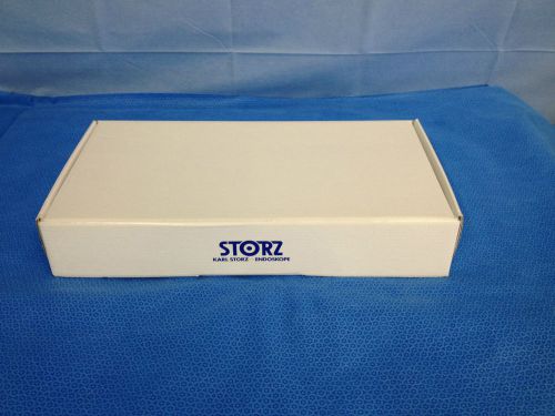
NEW IN BOX Karl Storz 20093801U1-DR SCB OR1 Control NEO System
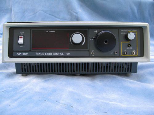
Karl Storz Xenon Light Source 611
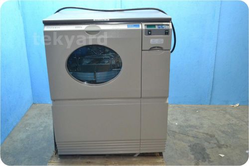
STERIS RELIANCE EPS ENDOSCOPE PROCESSING SYSTEM ! (125661)
Olympus LF-2 Flexible Intubation Scope / Fiberscope 4mm x 600mm (7482)
PENTAX FS-34P FLEXIBLE SIGMOIDOSCOPE (FIBER SCOPE) ! (132903)
PENTAX FG-34A ENDOSCOPE ! (132902)
By clicking "Accept All Cookies", you agree to the storing of cookies on your device to enhance site navigation, analyze site usage, and assist in our marketing efforts.
Accept All Cookies