US $1700
| Condition: |
Used: An item that has been used previously. The item may have some signs of cosmetic wear, but is fully
operational and functions as intended. This item may be a floor model or store return that has been used. See the seller’s listing for full details and description of any imperfections.
...
|
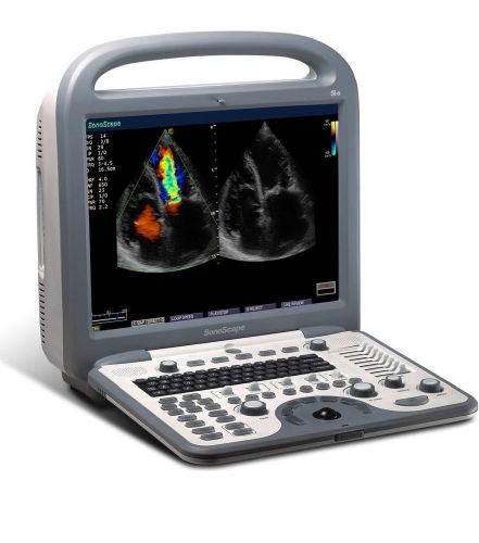
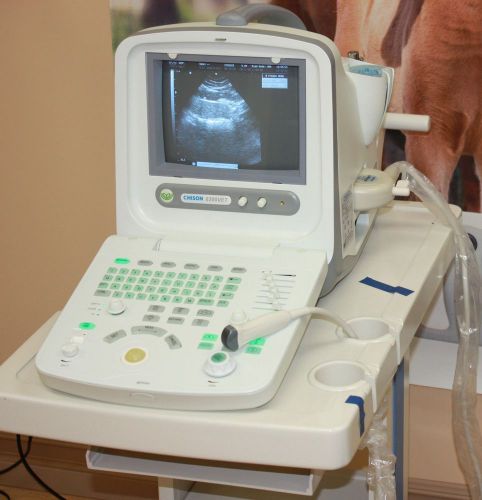
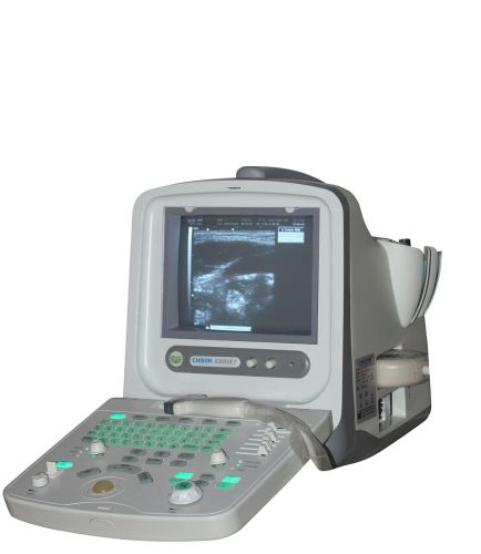
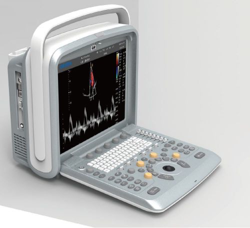
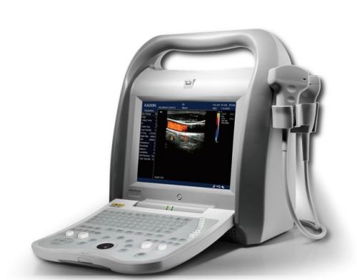
Portable ultrasound scanner | Cheap ultrasound scanner |
Directions
Similar products from Other Medical Equipment
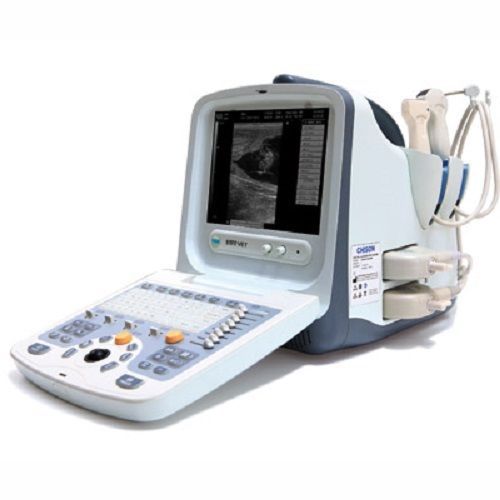
Chison 8300 Ultrasound machine &Linear array probe 5-10MHz&free deluxe trolley
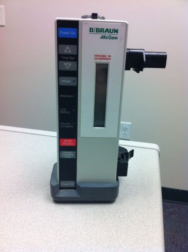
B BRAUN McGaw 360 Infuser Pump IV Infusion
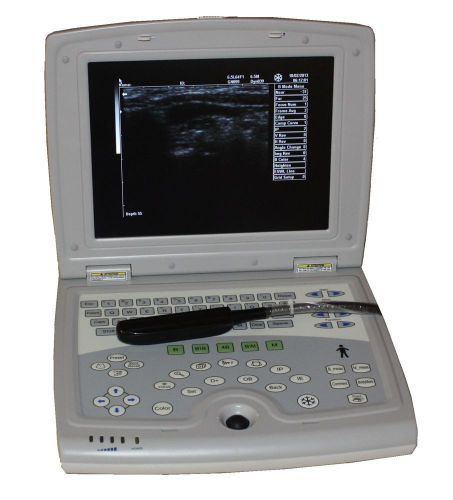
Amazing Veterinary Laptop ultrasound scanner LED 10"&rectal probe-USA warranty
Chison 8300Vet Veterinary Ultrasound Sacnner&Micro-Convex probe 5.0-8MHz-Demo
Chison 8300Vet Veterinary Ultrasound scanner &Linear array probe 5-10MHz-Demo
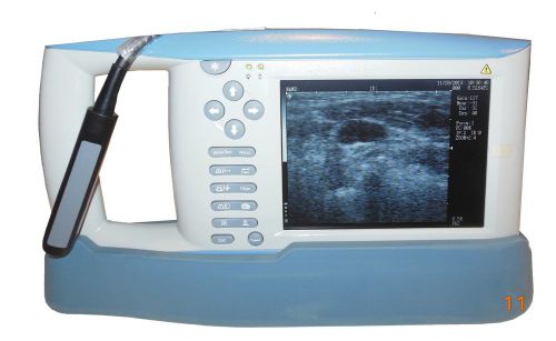
Veterinary Handheld Palm Ultrasound scanner,machine&rectal probe&Demo-USA seller
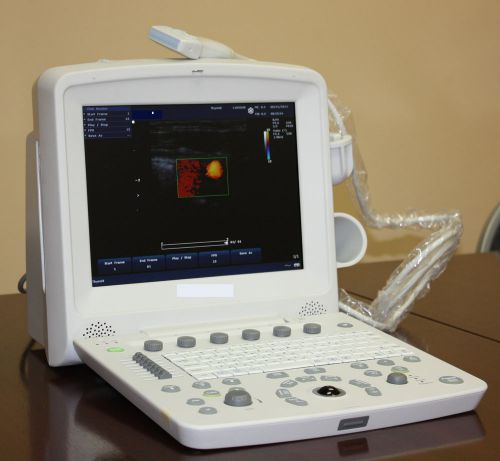
Most affordable Color Doppler Ultrasound Scanner,linear array probe FDA approved
Chison Q9Vet Veterinary Color Doppler Ultrasound Scanner&Micro convex probe Best
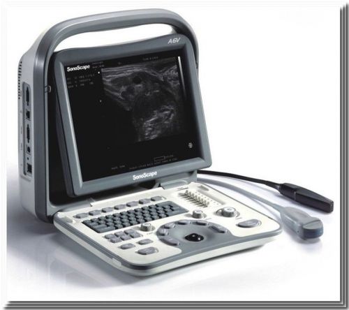
SonoScape A6V veterinary ultrasound scanner&rectal probe 5-12MHz-Demo model-Deal
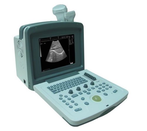
Veterinary digital ultrasound scanner machine-with rectal probe very stable-new
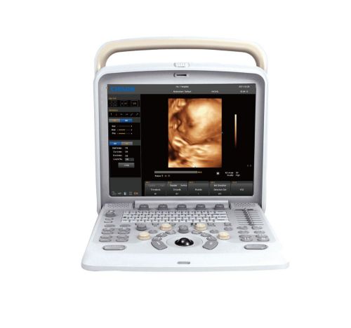
Chison Q5 Color Doppler Ultrasound Scanner& 4D Probe And Software Demo Model
Color Doppler Ultrasound Scanner&two probes FDA approved 12" screen-best deal

Kendall SCD Express with Vascular Refill Detection

Pistola dispensadora de capsulas de composite para odontologia.
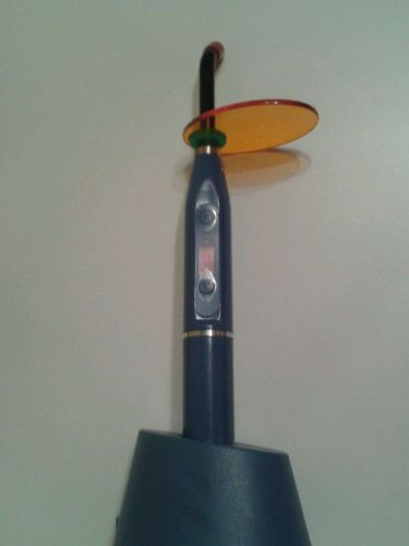
Lampara dental polimerizar led.
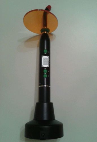
Lampara dental risingmed, led inalambrica.
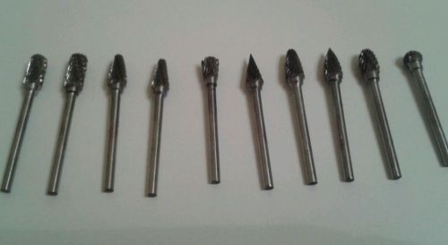
Fresas laboratorio vastago 3mm. Micromotor dental.
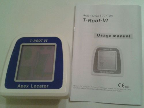
Localizador apices endodoncia dental modelo t-root 6. Nuevo. Con accesorios incl
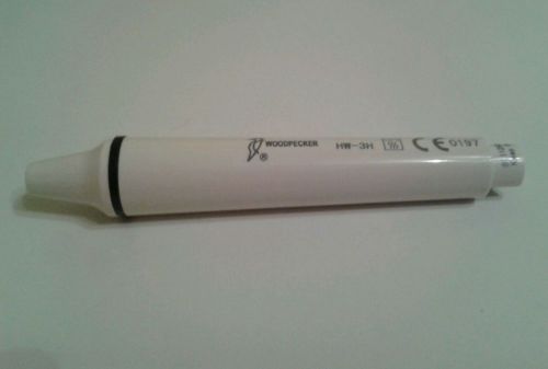
Terminal de ultrasonidos compatible con puntas y mangueras ems.
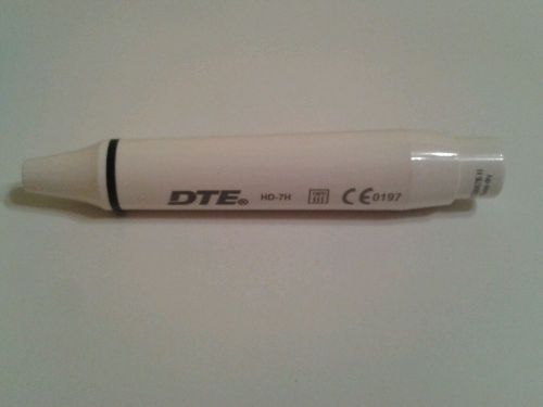
Terminal de ultrasonidos compatible con puntas y equipos satelec.
People who viewed this item also vieved
HP 21221A 1.9MHz Doppler Pencil Probe for HP Sonos 1000 to 4500 & 5500 (10521)
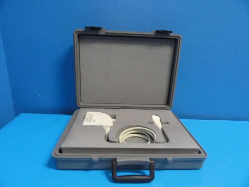
Siemens Omnia Versa 3.5P14 Phased Array 3.5MHz Transducer W/ Case (10340)
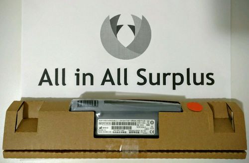
SONOSITE BATTERY FOR TITAN - MICROMAXX - M-TURBO REF: P07168-20
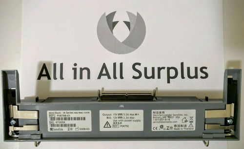
SONOSITE MINI DOCK FOR M-TURBO P08788-01
By clicking "Accept All Cookies", you agree to the storing of cookies on your device to enhance site navigation, analyze site usage, and assist in our marketing efforts.
Accept All Cookies