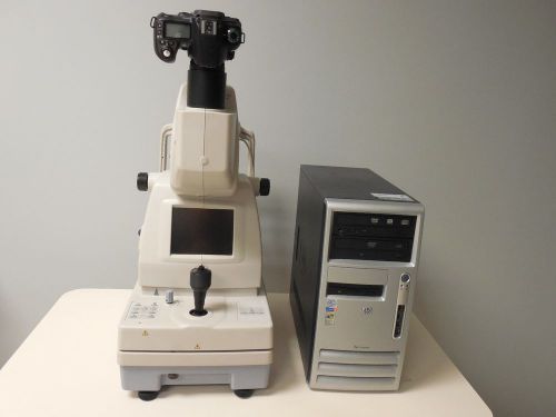
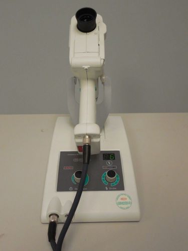
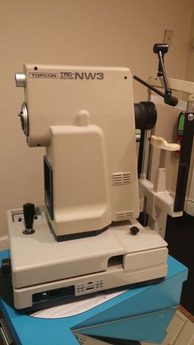
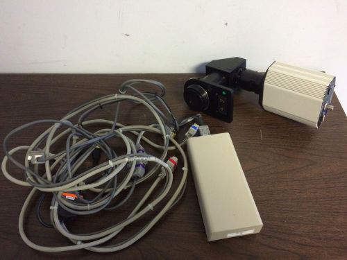
US $4,499.95
Directions
Similar products from Retinal Digital Cameras
Canon DGI Retinal Camera with Synamed Digital System
Topcon TRC NW100 Retinal Camera
Topcon NW6S Retinal Camera System
Kowa Genesis-d handheld retinal camera

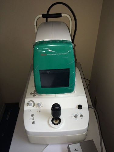
USED FUKUDA FF-3000 DIGITAL NM RETINAL CAMERA
Ophthalmic Imaging Systems (OIS) digital camera w/ adapter & Synch box 4 IX/EX
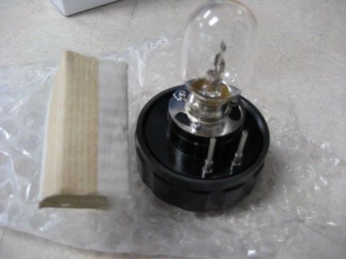
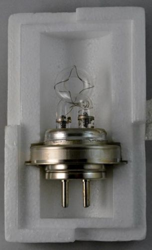
Brand new Topcon Illumination Bulb, part # 40524-19000. NO RESERVE.
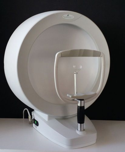
Medmont automated perimeter model: M700
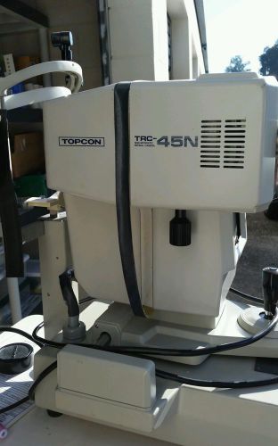
Topcon TRC-45N Polaroid Retinal Camera TRC45N
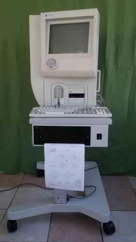
Zeiss Humphrey 740 HFA-II Visual Field Perimeter 14.0 Software.
Heidelberg HRT II Retinal Tomograph Glaucoma
TOPCON TRC NW200 Non Mydriatic Retinal/Fundus Camera
KOWA non-myd alpha-d Fundus Camera
Carl Zeiss Fundus Camera FF4 Fluorescein Pics. This Camera Is For CRA'S Only.
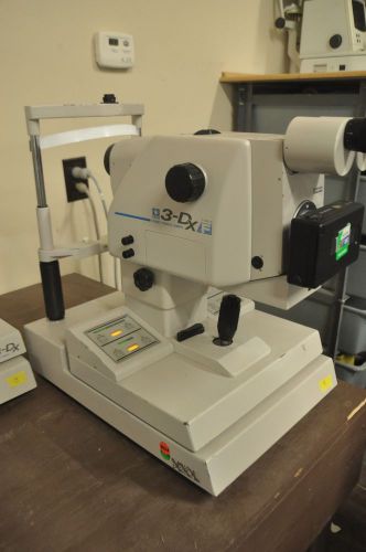
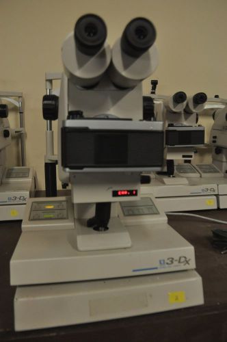
Nidek 3-Dx Type B Fundus Camera
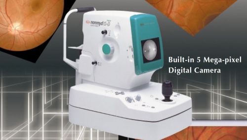
KOWA NONMYD ?-D DIGITAL NM RETINAL CAMERA WITH SOFTWARE NOTEBOOK WIN7
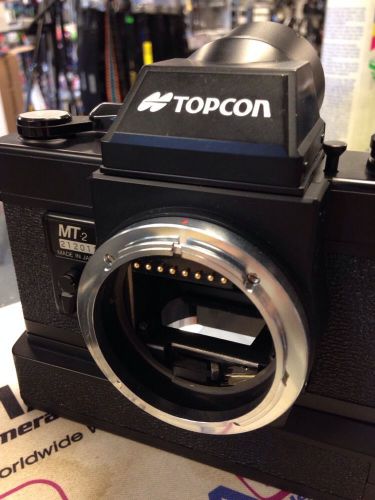
Strange Odd Cool Topcon MT-2 Fundus Camera for TRC-50X Mydriatic Photography
People who viewed this item also vieved
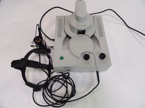
Coherent Lumenis LIO Surgical Laser Indirect Ophthalmoscope No Reserve!
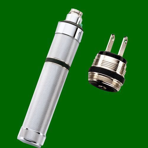
Welch Allyn 3.5v Direct Plug-in Rechargeable Handle - #AS20
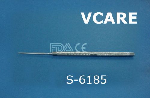
NEW Wecker Iris Spatula FDA & CE Approved
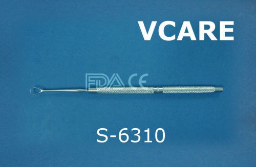
NEW Weber Lens Loop FDA & CE Approved
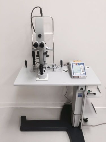
ZEISS VISULAS Yag II+ Plus Laser w Power Table & Slit Lamp
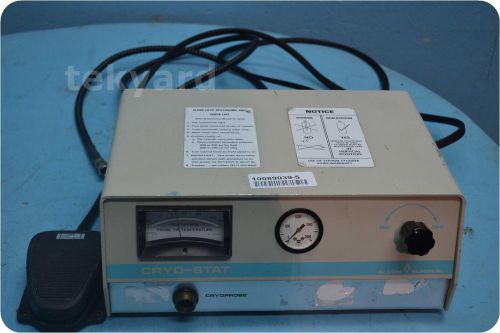
CRYOMEDICAL DNE-3000 CRYO-STAT / OPHTHALMIC CRYOSURGICAL UNIT @ (112916)
Volk 90D Diagnostic Surgical Lenses Indirect BIO Non Contact Lens MADE IN USA

20 D Lens Double Aspheric Lens And Case Optometry Equipment FG-87 Free Shipping
By clicking "Accept All Cookies", you agree to the storing of cookies on your device to enhance site navigation, analyze site usage, and assist in our marketing efforts.
Accept All Cookies